Cesarean Birth

The idea of delivering your baby by Cesarean can be totally frightening or overwhelming.  The birth of your sweet baby, who may have already died or who may have a condition not compatible with life, can already seem like just too much to bear.

You may either welcome, or dread, the knowledge that you will bear a scar of his or her delivery.  However you may feel about it, your feelings are OK.

To help make your Cesarean birth a little less overwhelming, it is a good idea to take a look at a Cesarean birth plan.

How far along are you?  Would you like to see the last developments of your baby?
Laparoscopic Surgery

Surgery for ectopic pregnancy may either be laparoscopy (explained here) or minilaparotomy.

Because ectopic pregnancy can be fatal to the mother unless the pregnancy ends as quickly as possible, I will only include very early development links to fetal information (and there is a probability that the development of an ectopic baby may be a little different; still, it can be nice to have a general idea of what your baby’s last developments will be). This surgical birth method may be used if methotrexate was ineffective.

The full medical term for laparoscopic surgery is “Laparascopic Salpingotomy”.  Laparoscopic surgery is performed under general anesthesia.  Your doctor will use a tool called a laparoscope to enter your abdomen through a small incision, deliver the baby, and to repair any affected part of the fallopian tube.

Once the doctor determines the condition of the fallopian tube, if it is not repairable, a “Laparoscopic Salpingectomy” will be performed (a “laparotomy”, which is a larger abdominal incision, may be required), which is the partial or the complete removal of the damaged fallopian tube.

You can make this birth method more meaningful by incorporating your own birth plan.

Development:

Blighted Ovum

A blighted ovum means that a fertilized egg has attached itself to your uterine wall, but the embryo (baby) did not develop. Cells developed to form the placenta and the amniotic sac, but not the embryo itself.

While a positive pregnancy test detects the placenta hormones (not an actual baby), finding out that you are pregnant can be the beginning of  hopes, aspirations and joy.

With a blighted ovum, your body may display signs of pregnancy, and may actually sustain the life of the growing placenta for a short time.  You may not know you have a blighted ovum until an ultrasound confirms it, or you may miscarry naturally before an ultrasound is performed.

The fact that a blighted ovum does not result in a baby can be equally–if not more–devestating than any other kind of miscarriage.

Finding out what to expect from your recommended birth method (listed below), and allowing yourself to experience healthy grief with a farewell celebration can be very useful and positive for you.

Please also utilize long term support services and emotional/spiritual health support services listed here in this website.

It is also very important to reach out, and tell others about your story.  Please consider sharing your experience with us here and reading the stories shared here by other mothers who’ve experienced loss through blighted ovum.

We’d be so honored to learn from you and to cry with you.

Birth Methods:

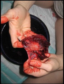
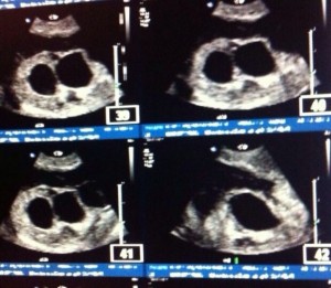
Below are photos of what you might expect to see or your blighted ovum to look like:

Triplet Blighted Ovum


Clicking the photo will direct you to its web source.

Clicking the photo will direct you to its web source.  You may find something similar to this during the course of your miscarriage.

Clicking the photo will direct you to its web source.  This is the sac, opened up.

Ectopic Pregnancy

An ectopic pregnancy means that your baby has attached itself to an area outside of the uterus rather than inside your actual uterus.  This situation can be fatal to the mother unless the pregnancy ends as quickly as possible.

This can be a very heartwrenching situation for a mother, who may mistakenly believe that she needs to have an “elective abortion”.  In an elective abortion, a mother electively chooses to terminate her pregnancy-despite the knowledge that the baby most probably would go on to develop through a full term pregnancy, and have a live birth.  In an ectopic pregnancy, the very high probability of both the baby dying and the mother dying, make delivering the baby as quickly as possible a necessity.

Nevertheless, we connect both mothers considering elective abortion and mothers facing ectopic pregnancy to our Maternal Death resources.  May you find the remainder of our ectopic pregnancy information below to be gentle during this impossibly difficult time.

How far along are you?  Because ectopic pregnancy can be fatal to the mother unless the pregnancy ends as quickly as possible, I will only include very early development links to fetal information (and there is a probability that the development of an ectopic baby may be a little different; still, it can be nice to have a general idea of what your baby’s last developments will be):

Birth Methods:

If your pregnancy is further along than this, you will probably give birth to your baby via laparoscopy surgery.

You are invited to share your story here as well: please remember that sharing your story at stillbirthday is a way to express your feelings and share your experiences with other mothers – it is not to diagnose, treat or answer any medical questions.

You might visit our farewell celebrations for ideas to celebrate your baby.

Molar Pregnancy

There are two types of molar pregnancy:

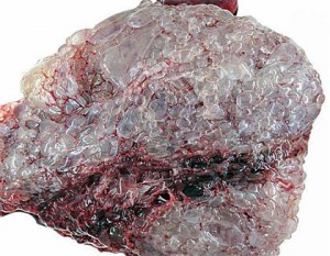
Complete molar pregnancy. An egg with no genetic information is fertilized by a sperm.  The sperm grows on its own, but it can only become a growth of placental tissue (hence a positive pregnancy test) and cannot become a fetus.  In a complete mole, all of the fertilized egg’s chromosomes (tiny thread-like structures in cells that carry genes) come from the father. Normally, half come from the father and half from the mother. In a complete mole, shortly after fertilization, the chromosomes from the mother’s egg are lost or inactivated, and those from the father are duplicated. As this tissue grows, it looks a bit like a cluster of grapes. This cluster of tissue can very rapidly fill the uterus.

Partial molar pregnancy. An egg is fertilized by two sperm.  If an abnormal embryo does begin to develop, it will quickly die because of the rapidly growing mass of abnormal tissue filling your uterus.  In most cases of partial mole, the mother’s 23 chromosomes remain, but there are two sets of chromosomes from the father (so the embryo has 69 chromosomes instead of the normal 46). This can happen when the chromosomes from the father are duplicated or if two sperm fertilize an egg.

Molar pregnancy poses a threat to the pregnant woman because it can occasionally result in a rare pregnancy-related form of cancer called choriocarcinoma (see end of document).


Molar pregnancy is assessed with a pelvic exam and ultrasound.  The abnormal placenta mass will have a clustered, grape like appearance.

For these and other serious medical risks, the molar pregnancy is immediately ended with medical support.  This is generally done with a D&C.  Afterward, you will have regular blood tests to look for signs of trophoblastic disease. These blood tests will be done over the next 6 to 12 months. Your doctor will caution you that you will need to use birth control for the next 6 to 12 months so that you don’t get pregnant. It is very important to see your doctor for all follow-up visits.

While a positive pregnancy test detects the placenta hormones (not an actual baby), finding out that you are pregnant can be the beginning of a hopes, aspirations and joy.

“The fact that a (complete) molar pregnancy does not result in a baby (or, twins) can be equally–if not more–devastating than any other kind of miscarriage.

Please be gentle on yourself and know that your loss is worthy to grieve.”

 – stillbirthday mother

Finding out what to expect from a D&C, and allowing yourself to experience healthy grief with a farewell celebration can be very useful and positive for you.

Please also utilize long term support services and emotional/spiritual health support services listed here in this website.

One stillbirthday mother suggests asking your trusted healthcare provider about drinking Chaga tea during your postpartum healing.

It is also very important to reach out, and tell others about your story.  Please consider sharing your experience with us here, and reading the stories shared here by other mothers who’ve experienced molar pregnancy.

We’d be so honored to learn from you and to cry with you.


~~~~~~~~~~

If you are interested in seeing what a molar pregnancy may look like,

below is a very graphic image:


~~~~~~~~~~

About Gestational Trophoblastic Disease – from cancer.org:

What is gestational trophoblastic disease?

Gestational trophoblastic disease (GTD) is a group of rare tumors that involve abnormal growth of cells inside a woman’s uterus. GTD does not develop from cells of the uterus like cervical cancer or endometrial (uterine lining) cancer do. Instead, these tumors start in the cells that would normally develop into the placenta during pregnancy. (The term gestationalrefers to pregnancy.)

GTD begins in the layer of cells called the trophoblast that normally surrounds an embryo. (Tropho- means nutrition, and -blast means bud or early developmental cell.) Early in normal development, the cells of the trophoblast form tiny, finger-like projections known as villi. The villi grow into the lining of the uterus. In time, the trophoblast layer develops into the placenta, the organ that protects and nourishes the growing fetus.

Most GTDs are benign (not cancer) and they don’t invade deeply into body tissues or spread to other parts of the body. But some are malignant (cancerous). Because not all of these tumors are cancerous, this group of tumors may be referred to as gestational trophoblastic disease, gestational trophoblastic tumors, or gestational trophoblastic neoplasia. (The word neoplasia simply means new growth.)

All forms of GTD can be treated. And in most cases the treatment produces a complete cure.

Types of gestational trophoblastic disease

The main types of gestational trophoblastic diseases are:

  • Hydatidiform mole (complete or partial)
  • Invasive mole
  • Choriocarcinoma
  • Placental-site trophoblastic tumor
  • Epithelioid trophoblastic tumor

Hydatidiform mole

The most common form of GTD is called a hydatidiform mole, also known as a molar pregnancy. It is made up of villi that have become swollen with fluid. The swollen villi grow in clusters that look like bunches of grapes. This is called a molar pregnancy, but it is not possible for a normal baby to form. Hydatidiform moles are not cancerous, but they can develop into cancerous GTDs.

There are 2 types of hydatidiform moles: complete and partial.

A complete hydatidiform mole most often develops when either 1 or 2 sperm cells fertilize an egg cell that contains no nucleus or DNA (an “empty” egg cell). All the genetic material comes from the father’s sperm cell. Therefore, there is no fetal tissue.

Surgery can totally remove most complete moles, but as many as 1 in 5 women will have some persistent molar tissue (see below). Most often this is an invasive mole, but rarely it is a choriocarcinoma, a malignant (cancerous) form of GTD. In either case it will require further treatment.

A partial hydatidiform mole develops when 2 sperm fertilize a normal egg. These tumors contain some fetal tissue, but this is often mixed in with the trophoblastic tissue. It is important to know that a viable (able to live) fetus is not being formed.

Partial moles are usually completely removed by surgery. Only a small number of women with partial moles need further treatment after initial surgery. Partial moles rarely develop into malignant GTD.

Persistent gestational trophoblastic disease is GTD that is not cured by initial surgery. Persistent GTD occurs when the hydatidiform mole has grown from the surface layer of the uterus into the muscle layer below (called the myometrium). The surgery used to treat a hydatidiform mole (called suction dilation and curettage, or D&C) scrapes the inside of the uterus. This removes only the inner layer of the uterus (the endometrium) and cannot remove tumor that has grown into the muscular layer.

Most cases of persistent GTD are invasive moles, but in rare cases they are choriocarcinomas or placental site trophoblastic tumors (see below).

Invasive mole

An invasive mole (formerly known as chorioadenoma destruens) is a hydatidiform mole that has grown into the muscle layer of the uterus. Invasive moles can develop from either complete or partial moles, but complete moles become invasive much more often than do partial moles. Invasive moles develop in a little less than 1 out of 5 women who have had a complete mole removed. The risk of developing an invasive mole in these women increases if:

  • There is a long time (more than 4 months) between the last menstrual period and treatment.
  • The uterus has become very large.
  • The woman is older than 40 years.
  • The woman has had GTD in the past.

Because these moles have grown into the uterine muscle layer, they aren’t completely removed during a D&C. Invasive moles can sometimes go away on their own, but most often more treatment is needed.

A tumor or mole that grows completely through the wall of the uterus may result in bleeding into the abdominal or pelvic cavity. This bleeding can be life threatening.

Sometimes after removing a complete hydatidiform mole, the tumor spreads (metastasizes) to other parts of the body, most often the lungs. This occurs about 4% of the time (or 1 in 25 cases).

Choriocarcinoma

Choriocarcinoma is a malignant form of GTD. It is much more likely than other types of GTD to grow quickly and spread to organs away from the uterus.

Choriocarcinoma most often develops from a complete hydatidiform mole, but it can also occur after a partial mole, a normal pregnancy, or a pregnancy that ends early (such as a miscarriage or an elective abortion).

Rarely, choriocarcinomas that are not related to pregnancy can develop. These can be found in areas other than the uterus, and can occur in both men and women. They may develop in the ovaries, testicles, chest, or abdomen. In these cases, choriocarcinoma is usually mixed with other types of cancer, forming a type of cancer called a mixed germ cell tumor.

These tumors are not considered to be gestational (related to pregnancy) and are not discussed in this document. Non-gestational choriocarcinoma can be less responsive to chemotherapy and may have a less favorable prognosis (outlook) than gestational choriocarcinoma. For more information about these tumors, see our documents, Ovarian Cancer and Testicular Cancer.

Placental-site trophoblastic tumor

Placental-site trophoblastic tumor (PSTT) is a very rare form of GTD that develops where the placenta attaches to the lining of the uterus. This tumor most often develops after a normal pregnancy or abortion, but it may also develop after a complete or partial mole is removed.

Most PSTTs do not spread to other sites in the body. But these tumors have a tendency to grow into (invade) the muscle layer of the uterus.

Most forms of GTD are very sensitive to chemotherapy drugs, but PSTTs are not. Instead, they are treated with surgery, aimed at completely removing disease.

Epithelioid trophoblastic tumor

Epithelioid trophoblastic tumor (ETT) is an extremely rare type of GTD that can be hard to diagnose. ETT used to be called atypical choriocarcinoma because the cells look like choriocarcinoma cells under the microscope, but it is now thought to be a separate disease. Because it can be found growing in the cervix, it can also sometimes be confused with cervical cancer. Like PSTT, ETT most often occurs after a full-term pregnancy, but it can take several years after the pregnancy for the ETT to occur. Also, like PSTT, ETT does not respond very well to chemotherapy drugs, so the main treatment is surgery. It might have already metastasized when it is diagnosed which carries a poorer prognosis (outlook).

You have permission to use this photo for social media awareness.  Just visit stillbirthday on Facebook to grab and share the photo.

Threatened Miscarriage

If your doctor told you that you are having a threatened miscarriage, you should know:

Many mothers with threatened miscarriage go on to have a complete pregnancy.

It is better to find and treat health problems (particularly systemic ones) before you get pregnant than to wait until you’re already pregnant.

Miscarriages are less likely if you receive early, comprehensive prenatal care and avoid environmental hazards such as x-rays, drugs and alcohol, high levels of caffeine, and infectious diseases.  Being obese or having uncontrolled diabetes might be considerations in an increase of risk for miscarriage.

The use of progesterone is controversial. It might relax smooth muscles, including the muscles of the uterus. However, it also might increase the risk of an incomplete miscarriage or an abnormal pregnancy. Unless there is a luteal phase defect, progesterone should not be used – please, consult the prospect of using any method of progesterone with your healthcare provider.  These might include Vitex Extract or Progesterone Crème.  Before the use of progesterone, your provider may offer Endometrin.

The use of false unicorn root (or other herbs such as cramp bark) is also controversial.  It is said to help “normalize” gynecological concerns with the uterus, including preventing miscarriage.  This native US herb is said to help facilitate the release of hormones by the ovaries.  Despite the claims to prevent miscarriage, there are warnings against using this herb in pregnancy.  Please consult with your medical provider before attempting to use any herbs or other non-medical resources to sustain your pregnancy.

You may be told to avoid or restrict some forms of activity. Not having sexual intercourse is usually recommended until the warning signs have disappeared.

Remember A+B+C = abdominal pain, bleeding, cramping.  These three together are signs of a probable miscarriage.

Questions for your provider can include the above as well as asking your provider to share their perspective and information regarding: beta HcG testing, progesterone level testing, antiphospholipids or anticardiolipids testing, and/or ultrasound.

Are you experiencing additional signs of miscarriage?


Additional suggestions later in pregnancy to avoid preterm labor and birth can include:

Our levels of augmentation page gives information regarding induction and augmentation, which you might contrast to the information on stalling or avoiding labor in any trimester.


Click here to go back to the different kinds of loss.

We also have information in our Getting Pregnant Again section that may prove helpful to you in this pregnancy – things that are encouraging, and other non-medical things you might consider.

You are invited to share your story here as well: please remember that sharing your story at stillbirthday is a way to express your feelings and share your experiences with other mothers – it is not to diagnose, treat or answer any medical questions.

Inevitable (or Incomplete) Miscarriage

An inevitable miscarriage is different from a threatened miscarriage, in that with an inevitable miscarriage, your baby will most certainly be born via miscarriage.

There are two situations that result in an inevitable (or incomplete) miscarriage:

  • Your cervical opening begins to dilate (open) and you are having vaginal bleeding (see our article on signs of miscarriage).  This means that your body is beginning to deliver your baby.
  • Your baby has not developed (stayed the same size) over a two week period.  Your baby’s heartrate may be slowing, or have completely stopped.

An inevitable miscarriage might be first discovered by ultrasound at a routine doctor appointment, or if you are experiencing possible symptoms of miscarriage you may visit your OB or your emergency room for confirmation.  The emergency room experience is often considered very unpleasant, but it may be needed.  If you visit your local emergency room, consider these tips:

  • let the staff know immediately that you believe you may be miscarrying
  • ask about their bereavement support, including staff and materials
  • ask if there is a women’s, laboring, or miscarriage room within the emergency room, or if you can be transferred to the labor and delivery level if that is what you’d prefer.  Once on the L&D level, ask for a room away from other mothers.
  • you may need to fill your bladder to help locate your baby on ultrasound.  Ask about drinking water, and curling on your side, rather than recieving a catheter.  If one is needed, ask about what to expect once it is removed (you may see some blood in your urine, and you may be sore for several hours or longer).
  • if you give birth to your baby in the emergency room, inquire of your personal options.  Visit our early pregnancy hospital birth plan for more details.  Understand navigating hospital policies, including genetic testing, returning your baby’s physical form back to you after any testing, and any other questions you have.

If your baby is younger than about 12 weeks gestation, you may be given three options for delivery:

If your baby is older than about 12 weeks gestation (about the beginning of the second trimester), you may be given these options for delivery:

You are invited to share your story here as well: please remember that sharing your story at stillbirthday is a way to express your feelings and share your experiences with other mothers – it is not to diagnose, treat or answer any medical questions.

You might visit our farewell celebrations for ideas to celebrate your baby.

Stillbirth

We have information and birth plans for several stillbirth situations:


You are invited to share your story here as well: please remember that sharing your story at stillbirthday is a way to express your feelings and share your experiences with other mothers – it is not to diagnose, treat or answer any medical questions.

You might visit our farewell celebrations for ideas to celebrate your baby.

Missed Miscarriage

If your doctor told you that your baby’s heart has stopped beating, you may be experiencing a missed miscarriage or an incomplete miscarriage.

You may have just found out that your baby’s heart actually stopped beating several days ago (or a couple of weeks ago) and you are just now beginning to see the earliest signs of delivery (see symptoms of a miscarriage for a complete listing, but includes seeing blood and/or pieces of tissue passing from your vagina).

A missed miscarriage occurs when your baby has already died, but the actual birthing process either has not yet begun or isn’t fully complete.

If your baby is younger than about 12 weeks gestation, you may be given three options for delivery:

If your baby is older than about 12 weeks gestation (about the beginning of the second trimester), you may be given these options for delivery:

You are invited to share your story here as well: please remember that sharing your story at stillbirthday is a way to express your feelings and share your experiences with other mothers – it is not to diagnose, treat or answer any medical questions.

You might visit our farewell celebrations for ideas to celebrate your baby.

Complete Miscarriage

This means that the baby has already been delivered, and the entire uterine lining and placenta have also been expelled.

This means that you are no longer pregnant.

If your pregnancy was very early, you can learn more about what happened from the natural miscarriage article.

Please visit these links for additional support:


The SBD® Doula provides support to families experiencing birth in any trimester and in any outcome.

Here at stillbirthday.info, you can learn about the SBD® Doula.